<

Research Projects

Stem Cell Biology

Stem cells continuously perceive and respond to various signals to maintain their biological fitness. Intestinal stem cells (ISCs), located at the vital barrier that separates the external and internal environments, integrates inputs from both outside and inside, including nutrients, microbes, cytokines, immunocytes, and neurons, to achieve tissue homeostasis. This makes the ISCs an excellent model of adult stem cell biology, tissue regeneration, and tumorigenesis. The Drosophila ISCs share many similarities with its mammalian counterpart, including general anatomical architectures, cellular physiology, cell lineages, and regulatory pathways, including the Notch, JNK, JAK/STAT, Insulin, Hippo, Wg, and EGF. These features make the discoveries in fly ISCs bear significant translational value for human physiology and pathology.


Recently, we discovered Piezo, a stretch-activated ion channel, as an important regulator of pre-EE (enteroendocrine) differentiation through the direct sensing of mechanical stimuli (Nature, 2018), which highly resemble Piezo’s function in human neural stem cells.


Currently, we are using RNAseq after FACS (fluorescence-activated cell sorting) to profile the pre-EE cells to get a comprehensive dataset about the signaling network underlying early EE determination. This dataset will provide the basis for further selective RNAi screens in the pre-EE population.


Visualize the Dynamics

Using new optical tools and techniques, we are aiming to discover and understand the constant changing biological system at the cellular and molecular level. Two major directions that are currently undertaken in our lab:

First, establish a new live imaging platform that allows us to observe the live tissues that previously difficult to study. With our latest ultrafast multicolor Thunder imaging system, we are planning to track multiple key molecules in complex developing organs with a high spatiotemporal resolution, in order to discover new behaviors, uncover the interactions between different signals, and explore the communications between cells and even organs.

Second, develop new probes to reveal the dynamic information in vivo. Previously, we generated a transcriptional timer that can encode the temporal information into a color ratio (eLife, 2019). This new tool led to a discovery of several new genes expressed in a subpopulation of fly intestinal stem cells, including the mechanosensitive channel Piezo (Nature, 2018).

Currently, we are developing new tools to tag the aged proteins and organelles to study their temporal dynamics in developing and aging tissues.

Organ Morphogenesis

As one of the three basic questions in developmental biology, studying morphogenesis not only provides the fundamental knowledge about life, but also pave the way for tissue engineering and regenerative medicine.  Our lab is interested in understanding how specific cytoskeleton component is spatially and temporally regulated in individual cell to dictate the global tissue shape.

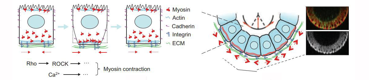
Previously, we identified a globally organized basal actomyosin contraction that promotes tissue elongation during fly oogenesis (Nat. Cell Bio. 2010).

And this global effect of actomyosin contraction can be revealed by acute removal of the basement membrane by collagenase (Mol. Bio. of the Cell, 2014).

Using this in vivo model, we are looking for new regulators that control the position and property of these contracting cells. We are also trying to identify the key switch that turn on this machinery, and test if the system can be engineered to create novel organ structures.  

Synthetic Biology/Genetic Tools


Traditional developmental biology is a “top-down” study that dissects the complex system into individual parts. However, information about how these biological parts connect to each other, and what are the minimal core components is usually missing. Meanwhile, many biological processes are hard to track due to the lack of a good read-out. To solve these problems, we take a “bottom-up” approach, which is to understand biology by reassembling the machinery and building de novo functions. With the development of the latest protein and genome engineering technology, we are now able to design novel molecules and test its effect in complex multicellular organisms such as the fly.

Previously, we designed a synthetic Notch receptor (Dev. Cell, 2015) and applied it in detecting direct cell-cell interaction as well as triggering tumor and wild-type cell competition in vivo (PNAS, 2017).

Now, we are directly modifying the endogenous signaling molecule to create synthetic bio-parts, which will allow us to build a new signaling circuit that can visualize and trigger the target biological activity.